Microscope Project: Onion Skin Abstracts
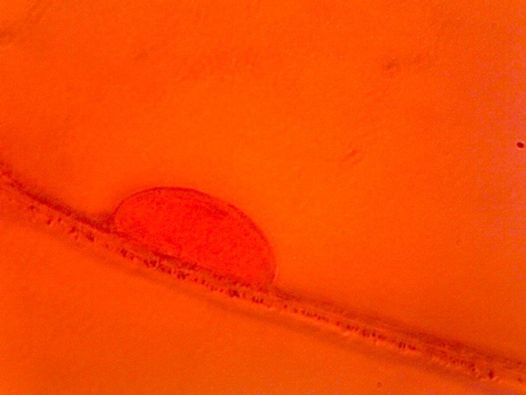
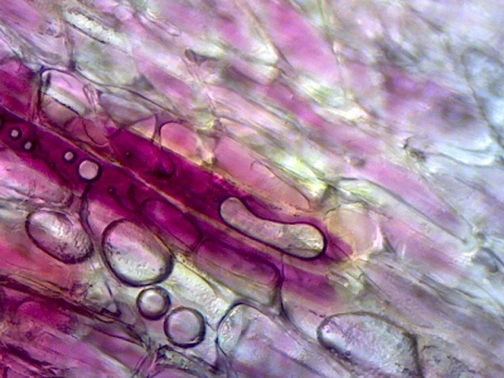
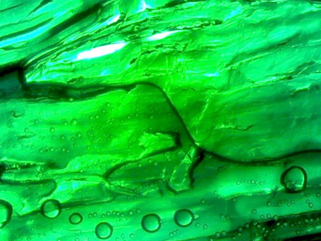
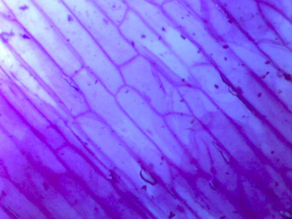
/I was preparing lunch yesterday - cutting up a red onion - when I realized that thin pieces of onion would be good subjects for the microscope. My goal was to do something artsy rather than scientific. See the results in the slide show below.
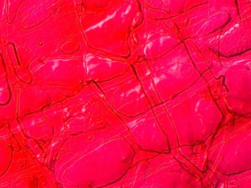
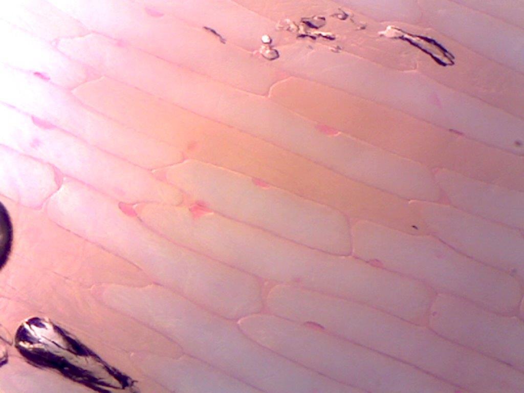
The outmost layer was very dry and stiff but I put a small piece on a slide. It looked like a brilliant pink stone wall under the microscope.
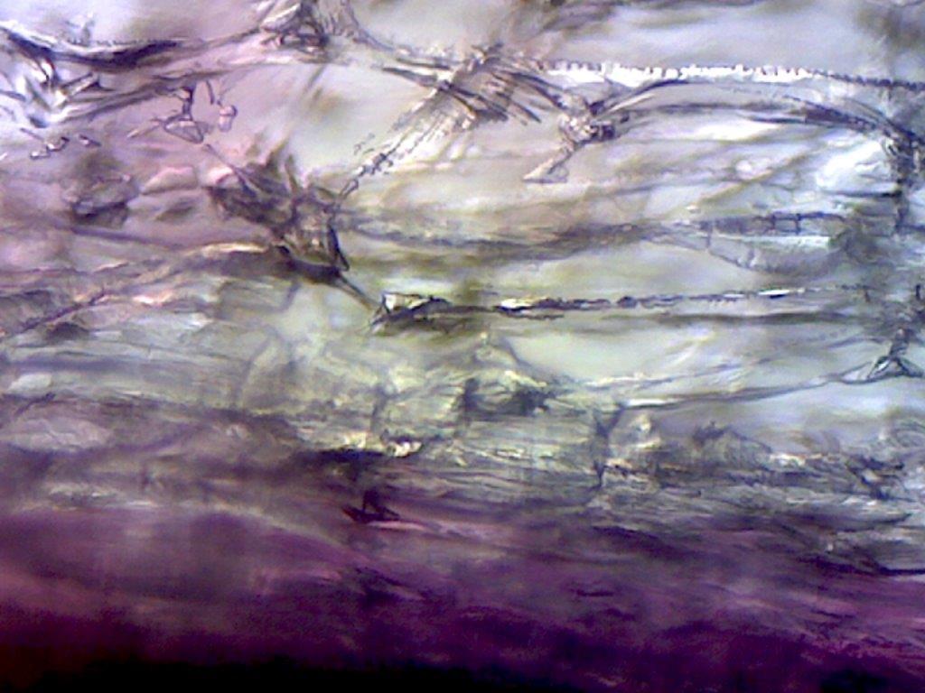
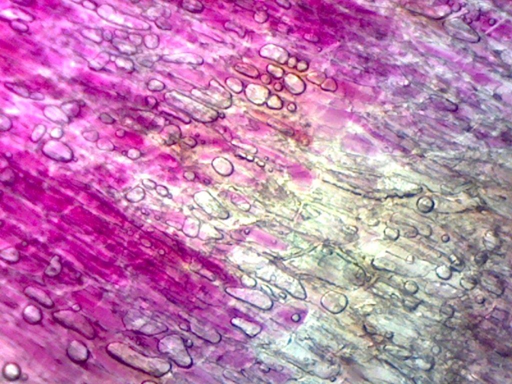
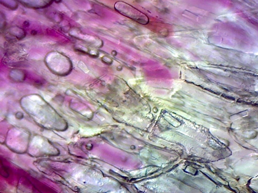
The next layer of the onion was more flexible but still not quite soft enough to be edible. I cut some small pieces. They looked various shades of deep purple, reddish and then clear. They dried out a bit before I got them under the microscope and I discovered that the bubbles made them just as interesting as the cell structures.
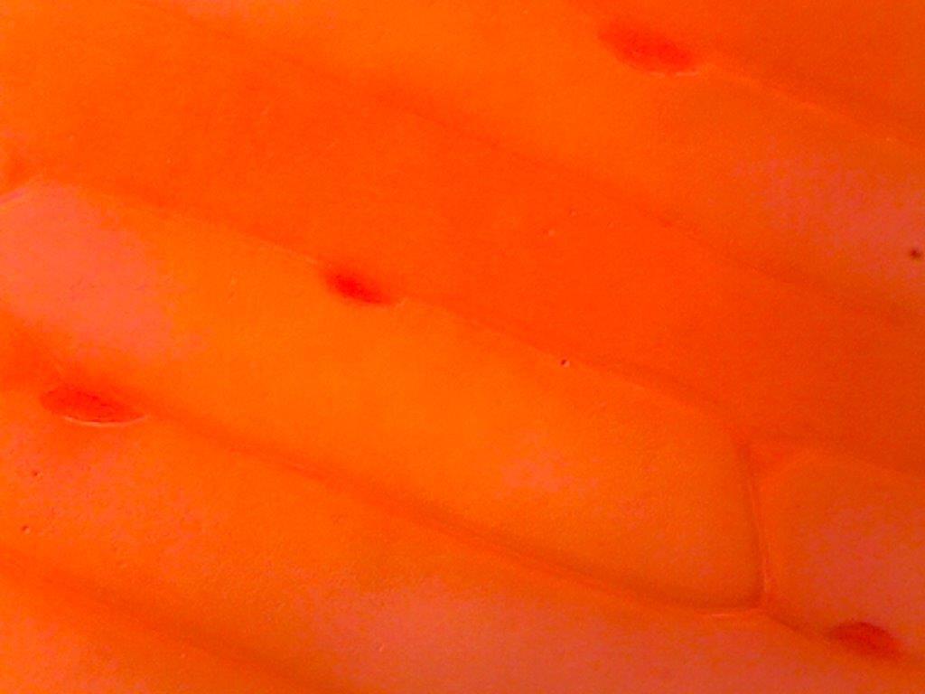
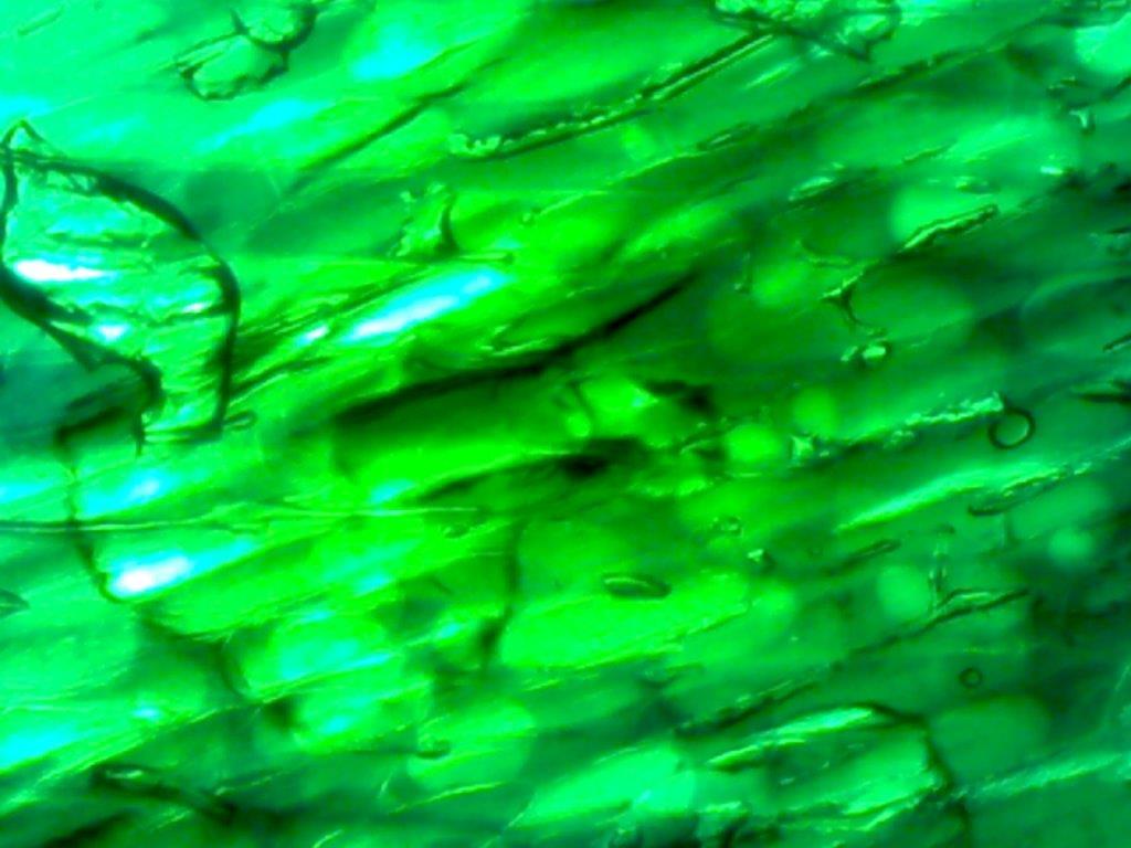
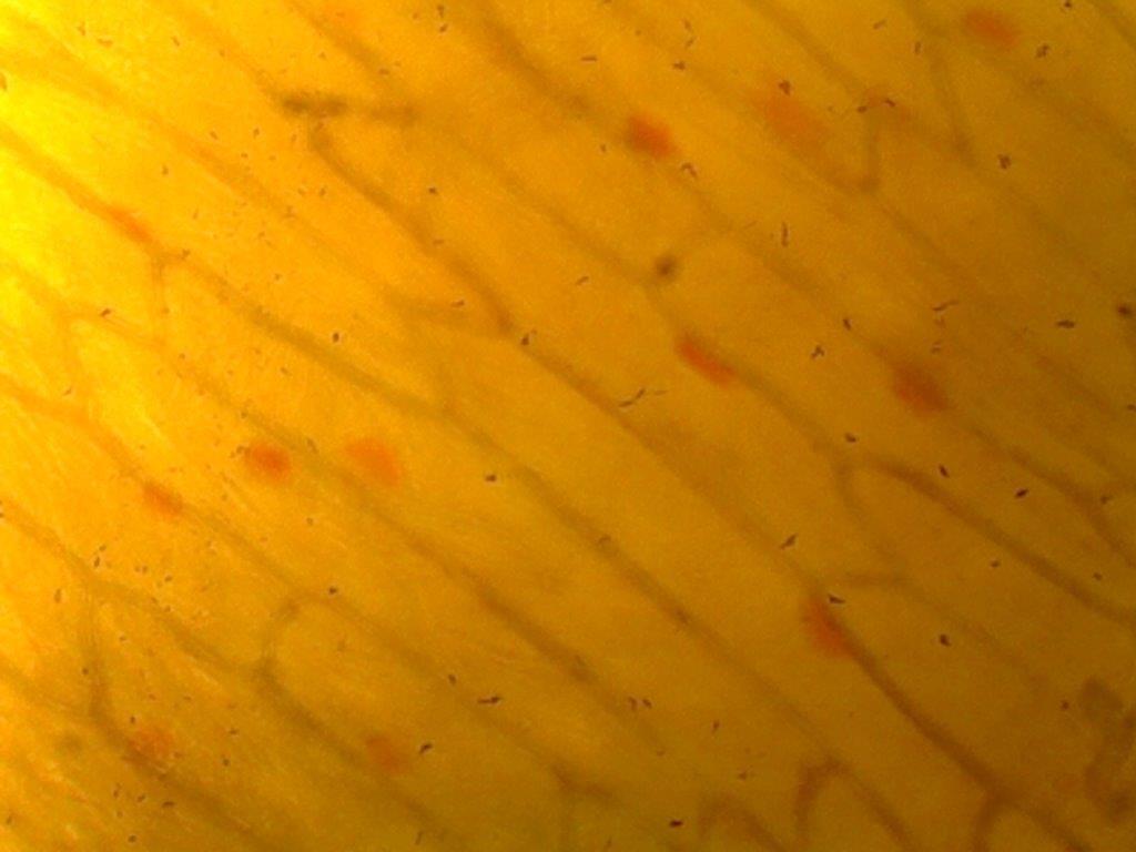
When I was dicing the onion - one of the major layers fell away and a think membrane - without color - was visible. I put several pieces of it on slides and tried some dyes/food coloring to get the green, yellow, orange, and purple images.

